
Congratulations to the REACH investigators listed below for their recent published work!
- BACPAC REACH
- Apr 1, 2022
- 2 min read
Dr. Anastasia Keller and Abel Torres-Espin et al. published paper "Unsupervised machine learning on motion capture data uncovers movement strategies in low back pain," which identified divergent sit-to-stand (STS) movement strategies in two groups of patients with cLBP vs. healthy individuals. Increased spinal loading, associated with the inefficient leaning STS strategy observed in patients with cLBP may underly the low back pain chronicity. Overall, the leaning STS strategy appears counterintuitive to the LBP condition, pointing to the underlying extensor muscle weakness of the primary movers (knee/hip extensors) during STS in patients with LBP. Data from this study suggests that cLBP patient physical therapy-based rehabilitation efforts should include lower extremity strengthening with the goal to correct biomechanical insufficiency identified in patients with LBP in the current work.
Read the abstract in Frontiers in Bioengineering and Biotechnology Journal here.

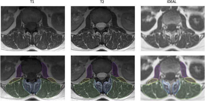
Another published manuscript by Dr. Nico Sollmann et al. titled "Paraspinal muscle in chronic low back pain: Comparison between standard parameters and chemical shift encoding-based water-fat MRI" investigated the ability of conventional MRI sequences to evaluate paraspinal muscle health compared with advanced MRI sequences. In general, assessments of muscle health with conventional MRI compared favorably to advanced MRI, although the advanced MRI sequences were able to detect early/subtle changes in muscle health not detectable with the conventional sequences.
Read the full article in Journal of Magnetic Resonance Imaging here.

Graphic 1: Goutallier classification for paraspinal musculature on conventional T1-weighted MRI images. Goutallier Grade: 0—normal muscle (a); 1—some fatty streaks (b); 2—less than 50% fatty muscle atrophy (c); 3—50% fatty muscle atrophy (d); and 4—greater than 50% fatty muscle atrophy (e).


Comments